Most patients that present with hip problems in young adulthood suffered damage to the hip during childhood due to either developmental dysplasia of the hip, perthes disease or slipped upper femoral epiphysis. All of these conditions can lead to early degeneration of the structures of the hip and associated pain and joint destruction. Some patients may have been diagnosed and treated by paediatric orthopaedic surgeons, but often milder forms may go unnoticed and are only diagnosed in early adulthood.
SLIPPED UPPER FEMORAL EPIPHYSIS
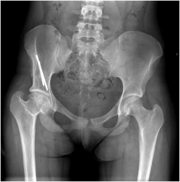
Slipped Upper Femoral Epiphysis (SUFE for short) occurs in the early teenage years and is due to the top of the ball of the hip joint (epiphysis) slipping off the top of the femur through the growth plate (physis). The result is that the hip joint becomes mis-shaped and predisposes the hip joint to early arthritis and damage to the soft tissues around the hip (the labrum). This usually starts as femoro-acetabular impingement but culminates in hip arthritis.
FEMORACETABULAR IMPINGEMENT (FAI)
Femoracetabular impingement describes the phenomenon where an abnormality of the shape of either the acetabulum (socket) or proximal femur (ball) leads to abnormal contact between the two bones that comprise the hip joint. In addition the soft tissues around the hip (labrum) can become damaged (labral tear) and this can cause pain and lead to early arthritis.
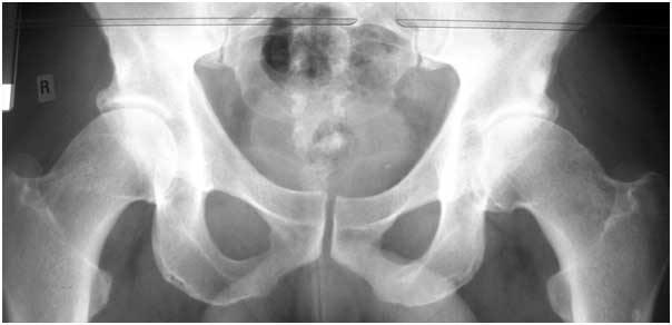
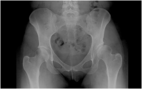
The diagnosis is usually made through consultation and examination with xrays that demonstrate the typical appearance of either a “cam” deformity of the femur (ball) or “pincer” deformity of the acetabulum (socket). In addition an MRI scan is usually required to evaluate the soft tissue that surround the hip joint most notably the labral cartilage which is often torn due to the bones pinching the labrum repetitively.
Once the diagnosis is established treatment may include hip injections, physiotherapy, pain relief or anti-inflammatories. In early FAI without significant arthritis some patients may benefit from key-hole surgery (hip arthroscopy) but where damage has progressed too far hip replacement surgery may be the only solution.
PERTHES DISEASE
Perthes disease (Legg-Calve-Perthes) occurs due to disruption of the blood supply to the hip in childhood most commonly in boys between the ages of 4 and 8 years. This is very similar to avascular necrosis in adults but children have wonderful healing ability and in 60% of cases go on to resolve without the need for any surgery. Interestingly, the prognosis is better in younger children (<6 years) where they have longer for their bones to heal and regain their normal shape.
Interruption of the blood supply of the femoral head leads to resorption of bone, followed by fragmentation, re-ossification and finally remodelling of the femoral head over a period of years. Where the head returns to the normal spherical shape the prognosis is excellent with a low likelihood of hip arthritis. Occasionally significant deformity does occur and may result in the need for surgery during both childhood or young adulthood.
Surgery for perthes may include osteotomies of the femur or pelvis (braking the bones and restting them) to try and optimise the remaining life of the hip joint. Where the destruction is too advanced to allow symptomatic relief complex total hip replacement surgery may be undertaken.
DEVELOPMENTAL DYSPLASIA OF THE HIP
Developmental dysplasia of the hip(DDH) occurs as a result of the abnormal development of the hip socket and results in a subluxed (partially dislocated) or dislocated hip (where the ball comes out of the socet) in infancy. It’s a complex condition with a number of risk factors which are screened for in the UK such as breach presentation, oligohydramnios (too little amniotic fluid in utero), family history and packaging deformities (due to the position the baby developed in utero).
Infants are routinely screened in the UK when they have their baby checks in the post-natal period, and may be referred for ultrasound for diagnosis.
The treatment of diagnosed DDH was revolutionised with the introduction of the Pavlik harness which is a simple device that positions the hip in the correct position for the hip to mature properly into a normal ball and socket joint. Successful treatment can be achieved in 90% of cases. The remaining 10% of cases will often require surgery in infancy or early childhood and a large number of procedures are described that involve putting the hip back in joint and keeping it in that position by braking and re-setting the bones.
Despite the very best care in childhood a proportion of patients will go on to require surgery in adulthood as the result of an abnormally formed hip. The surgeries may involve braking and resetting the bones (peri-acetabular or femoral osteotomy) where there is still some chance of preserving the native hip joint. Where too much damage has occurred often the best option to restore function and relieve pain is to proceed to hip replacement surgery. This is not without the risk of requiring revision hip surgery in later life, but technology is evolving and with it the survivorship of hip replacement prostheses is expected to increase.


